

Scientists map part of a mouse’s brain that’s so complex it looks like a galaxy
Advertisement
Read this article for free:
or
Already have an account? Log in here »
To continue reading, please subscribe:
Monthly Digital Subscription
$1 per week for 24 weeks*
- Enjoy unlimited reading on winnipegfreepress.com
- Read the E-Edition, our digital replica newspaper
- Access News Break, our award-winning app
- Play interactive puzzles
*Billed as $4.00 plus GST every four weeks. After 24 weeks, price increases to the regular rate of $19.95 plus GST every four weeks. Offer available to new and qualified returning subscribers only. Cancel any time.
Monthly Digital Subscription
$4.99/week*
- Enjoy unlimited reading on winnipegfreepress.com
- Read the E-Edition, our digital replica newspaper
- Access News Break, our award-winning app
- Play interactive puzzles
*Billed as $19.95 plus GST every four weeks. Cancel any time.
To continue reading, please subscribe:
Add Free Press access to your Brandon Sun subscription for only an additional
$1 for the first 4 weeks*
*Your next subscription payment will increase by $1.00 and you will be charged $16.99 plus GST for four weeks. After four weeks, your payment will increase to $23.99 plus GST every four weeks.
Read unlimited articles for free today:
or
Already have an account? Log in here »
Hey there, time traveller!
This article was published 09/04/2025 (354 days ago), so information in it may no longer be current.
WASHINGTON (AP) — Thanks to a mouse watching clips from “The Matrix,” scientists have created the largest functional map of a brain to date – a diagram of the wiring connecting 84,000 neurons as they fire off messages.
Using a piece of that mouse’s brain about the size of a poppy seed, the researchers identified those neurons and traced how they communicated via branch-like fibers through a surprising 500 million junctions called synapses.
The massive dataset, published Wednesday by the journal Nature, marks a step toward unraveling the mystery of how our brains work. The data, assembled in a 3D reconstruction colored to delineate different brain circuitry, is open to scientists worldwide for additional research – and for the simply curious to take a peek.
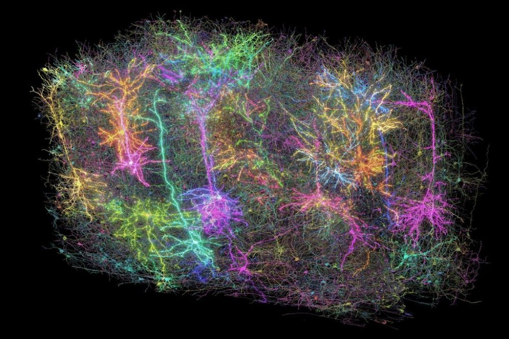
“It definitely inspires a sense of awe, just like looking at pictures of the galaxies,” said Forrest Collman of the Allen Institute for Brain Science in Seattle, one of the project’s leading researchers. “You get a sense of how complicated you are. We’re looking at one tiny part … of a mouse’s brain and the beauty and complexity that you can see in these actual neurons and the hundreds of millions of connections between them.”
How we think, feel, see, talk and move are due to neurons, or nerve cells, in the brain – how they’re activated and send messages to each other. Scientists have long known those signals move from one neuron along fibers called axons and dendrites, using synapses to jump to the next neuron. But there’s less known about the networks of neurons that perform certain tasks and how disruptions of that wiring could play a role in Alzheimer’s, autism or other disorders.
“You can make a thousand hypotheses about how brain cells might do their job but you can’t test those hypotheses unless you know perhaps the most fundamental thing – how are those cells wired together,” said Allen Institute scientist Clay Reid, who helped pioneer electron microscopy to study neural connections.
With the new project, a global team of more than 150 researchers mapped neural connections that Collman compares to tangled pieces of spaghetti winding through part of the mouse brain responsible for vision.
The first step: Show a mouse video snippets of sci-fi movies, sports, animation and nature.
A team at Baylor College of Medicine did just that, using a mouse engineered with a gene that makes its neurons glow when they’re active. The researchers used a laser-powered microscope to record how individual cells in the animal’s visual cortex lit up as they processed the images flashing by.
Next, scientists at the Allen Institute analyzed that small piece of brain tissue, using a special tool to shave it into more than 25,000 layers, each far thinner than a human hair. With electron microscopes, they took nearly 100 million high-resolution images of those sections, illuminating those spaghetti-like fibers and painstakingly reassembling the data in 3D.
Finally, Princeton University scientists used artificial intelligence to trace all that wiring and “paint each of the individual wires a different color so that we can identify them individually,” Collman explained.
They estimated that microscopic wiring, if laid out, would measure more than 3 miles (5 kilometers). Importantly, matching up all that anatomy with the activity in the mouse’s brain as it watched movies allowed researchers to trace how the circuitry worked.
The Princeton researchers also created digital 3D copies of the data that other scientists can use in developing new studies.
Could this kind of mapping help scientists eventually find treatments for brain diseases? The researchers call it a foundational step, like how the Human Genome Project that provided the first gene mapping eventually led to gene-based treatments. Mapping a full mouse brain is one next goal.
“The technologies developed by this project will give us our first chance to really identify some kind of abnormal pattern of connectivity that gives rise to a disorder,” another of the project’s leading researchers, Princeton neuroscientist and computer scientist Sebastian Seung, said in a statement.
The work “marks a major leap forwards and offers an invaluable community resource for future discoveries,” wrote Harvard neuroscientists Mariela Petkova and Gregor Schuhknecht, who weren’t involved in the project.
The huge and publicly shared data “will help to unravel the complex neural networks underlying cognition and behavior,” they added.
The Machine Intelligence from Cortical Networks, or MICrONS, consortium was funded by the National Institutes of Health’s BRAIN Initiative and IARPA, the Intelligence Advanced Research Projects Activity.
—-
The Associated Press Health and Science Department receives support from the Howard Hughes Medical Institute’s Science and Educational Media Group and the Robert Wood Johnson Foundation. The AP is solely responsible for all content.